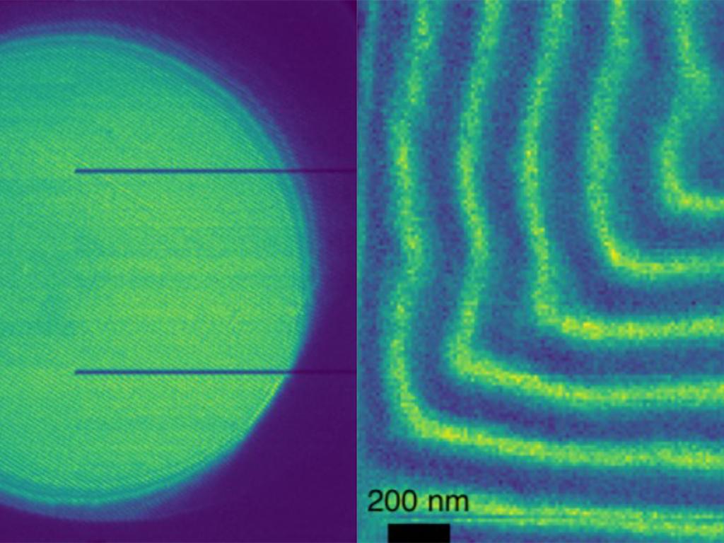
The University of Oregon’s Titan transmission electron microscope (TEM) system is a powerful tool for research in material science, electrochemistry, quantum physics, geosciences, and more. It is also widely used across the UO’s leading applied science programs, such as the Advanced Materials Analysis and Characterization graduate program, the Knight Campus Graduate Internship Program, and the planned Master’s in Quantum Engineering to be launched in fall 2024.
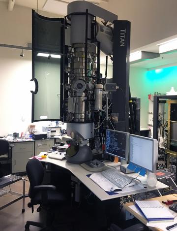
Researchers recently received awards from the National Science Foundation Major Research Instrumentation (NSF MRI) program and the Murdock Foundation to build out the capabilities of the Titan TEM, which funded the acquisition of a direct electron detector (MRI) and new electron energy loss spectrometer (Murdock). Researchers at the UO and Oregon State University (OSU) are currently pursuing funding to replace the existing Titan with a new, aberration-corrected scanning transmission electron microscope (STEM), which would be the first of its kind in the state of Oregon.
These new additions and enhanced capacity of the TEM lab at CAMCOR will accelerate several research projects in electron microscopy (Kayla Nguyen and Ben McMorran labs at the UO) and materials characterization (David Johnson and Carl Brozek labs at the UO and Melissa Santala’s group at OSU), to name a few.
The TEM is a critical asset for the Pacific Northwest. Since just 2020, 13 research groups representing the four major universities in the state of Oregon (UO, OSU, Portland State University, and Oregon Health & Science University) have all used the TEM to collect data to further their research fields. In addition to in-state usage, the TEM has been used by universities outside of the state, including the University of Wyoming and the University of Alberta.
Finally, the TEM is used as a training tool for multiple different degree programs. Since 2013, several undergraduates, 221 master’s students, and 58 PhD students have been trained to use the TEM. An additional 88 PhD students have used data collected by TEM facility director Josh Razink in their dissertations. The tool is also used for NSF Research Experiences for Undergraduates programs, as well as science outreach programs for state and local high schools.
The Titan TEM has not only been useful to university researchers, but government labs and private industry as well. One hundred and twenty researchers at more than 60 organizations have used the instrument since 2013. These organizations range from small start-up companies to corporations listed in the S&P 500, hailing from many different sectors, including the semiconductor and defense industries.