
Before new medical treatments or technologies can be developed, scientists first need to understand the basic biology behind them. At the University of Oregon, researchers are pursuing those questions with support from the National Institutes of Health’s (NIH) Maximizing Investigators’ Research Award (MIRA) program.
Funded through the NIH’s National Institute of General Medical Sciences (NIGMS), MIRA supports a researcher’s broader scientific program rather than a single, narrowly defined project. Awards generally provide five years of funding, giving researchers more stability to pursue ambitious work and adjust their approach as new questions and discoveries come up.
Most NIH research funding is organized around individual projects, with researchers proposing specific questions and experiments they plan to carry out. MIRA works a little differently. By supporting an investigator’s long-term research goals that align with the NIGMS mission, the program gives scientists more room to follow promising leads, including ones they may not have anticipated when they first applied for funding.
That flexibility can also provide more stability for research teams. With several years of support, investigators can plan farther ahead, invest in new technologies, and support students and other researchers working in their labs. At the UO, MIRA funding is supporting researchers working on a range of fundamental biological questions. Their research questions are different, but each is using the flexibility of the program to better understand some of the basic processes that shape life and health.
Genetic Long Division: Understanding the Secrets of How Cells Divide
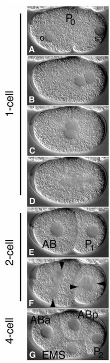
Every living thing begins as a cell that divides and divides again. For a creature to continue to grow and develop, its cells must divide correctly. Bruce Bowerman, biology professor and member of the Institute of Molecular Biology, is using the tiny roundworm Caenorhabditis elegans (better known as C. elegans or nematode worms) to better understand what happens inside cells during the division process.
With support from MIRA, Bowerman’s lab is studying the cytoskeleton, a network of proteins inside cells that helps them maintain their shape, move, and divide. His team is particularly interested in the earliest stages of development and how cells organize themselves during the division of egg cells and early embryos.
The researchers will combine genetic tools with advanced microscopes that let them watch these processes unfold in living cells. Technologies, including CRISPR gene editing, will allow the team to manipulate specific genes and look more closely at the physical properties of cells. One focus is how cells organize and separate their genetic material during division. Bowerman’s team is also looking at how structures inside a cell interact with its outer surface, and how those interactions help cells divide the way they should.
The research may focus on a microscopic worm, but the underlying questions are much broader. Many of the cellular processes Bowerman studies are shared across animals. Understanding them could provide insights relevant to human fertility, cancer research, tissue engineering, and other areas of biology.
It Takes Two to Tango: How Genetics Affect Infection and the Resulting Immune Response
When a bacterial infection occurs, there are two sets of genetics at play: those of the bacteria and those of the person they infect. Matthew Barber, associate professor of biology and member of the Institute of Ecology and Evolution, is studying how those differences can shape infection and how bacteria evolve in response to their hosts defenses.
Barber’s MIRA-funded research brings together evolutionary genetics and laboratory experiments to understand how bacterial pathogens and their hosts adapt to one another. His team is focusing on bacteria that cause human disease, including pathogens that are becoming increasingly resistant to antibiotics.
Part of the research examines proteins found on the surfaces of host and bacterial cells. Differences in these proteins can affect whether bacteria are able to enter and colonize the body, and if they do manage to break in, whether they can evade the immune system.
Barber’s team is also studying the antimicrobial enzymes that hosts produce to fight bacteria, along with the ways pathogens evolve to resist those defenses.
Combining genetic analysis with molecular biology and microbiology will allow the researchers to identify some of the genetic changes that can make an infection more or less successful. Over time, a clearer picture of these host-pathogen interactions could help researchers better understand why certain infections take hold and potentially point toward new ways of preventing or treating bacterial disease.
Go with the Flow: Investigating the Physical Forces Inside Cells
Daniel Grimes, associate professor of biology and member of the Institute of Molecular Biology, is studying how cells use physical forces, including tiny fluid flows, to communicate during development and maintain healthy tissues.
Grimes’ MIRA-funded research uses a zebrafish model to investigate how those flows are created and sensed by cilia—the tiny, hair-like structures that move fluid around cells.
His team will look at how flow signals help shape the developing body, including the role of the Reissner fiber, a thin, thread-like structure in the spinal canal that helps regulate body shape. They will also investigate proteins inside cilia that could provide new clues about how these structures work and how cells sense the movement of fluid around them.
Problems with these signaling processes have been linked to birth defects, spinal disorders, and ciliopathies. By looking closely at how cells sense and respond to physical forces, Grimes’ lab hopes to fill in some of the gaps in scientists’ understanding of how a healthy body takes shape in the first place.
Building on Biological Discovery
Taken together, the three projects show the range of questions that MIRA funding is helping UO researchers explore. Bowerman is looking at what happens as cells divide, Barber at the ongoing evolutionary back-and-forth between bacteria and their hosts, and Grimes at the physical signals that help organize a developing body.
There is not necessarily a straight line from this kind of fundamental research to a new treatment or technology. Often, that is not the immediate goal. Instead, researchers are working to understand the biological processes that later discoveries can build on. MIRA gives researchers more time and flexibility to do that work. For Bowerman, Barber, and Grimes, sustained NIH support means their labs can follow unexpected results, try new approaches, and keep asking questions as the science develops.