Research Revealed is sponsored by the Office of the Vice President for Research and Innovation and the Undergraduate Research Opportunity Program.
Contest Details
Important Dates
Contest opens February, 16, 2026.
Contest closes March, 13, 2026. Deadline extended to Friday, March 20, 2026.
Entrants will be notified by April 3, 2026.
Award winning photos will be displayed at the Week of Research and Innovation on April 7, 2026 and the Undergraduate Research Symposium on May 7, 2026.
Eligibility
The contest is open to all currently enrolled undergraduates at the University of Oregon.
One entry will be accepted per person.
Evaluation Criteria
Submissions may depict the subject of your research or capture the process of doing research. Entries will be judged on the following criteria:
Aesthetic appeal.
Creativity, composition, and visual impact.
Conciseness and clarity of the written description of the image.
Prizes
Prizes will be awarded in both the photo and multimedia categories.
First place: $250
Second place: $150
Third place: $100
One $50 people's choice winner will be selected during the Week of Research and Innovation.
University fiscal policy requires that monetary awards in excess of $50 are reported as taxable income to the recipient.
Submission Guidelines
Images of research in the humanities are encouraged. Photo by Lauryn Cole.
Size: Files must be no larger than 100 MB
Videos larger than this should be shared via a link to the video
Accepted formats
Image: .jpg, .png., or .tif
Minimum resolution: 300 dpi, vertical or horizontal orientations are accepted
Videos: .avi, .mp4, .mov
Audio: .mp3, .aif, .iff
Description of image or multimedia product (maximum 150 words): If the product features someone conducting research, be sure to describe what the person featured is doing. The description should be written so that someone who is not a researcher in your field can understand what is occurring in the photo, video, etc. The description is weighed heavily in determining winning entries.
If an individual appears in the image or video, a model release form must be completed for that person.
Images or videos containing human research subjects MUST have PI approval before submission.
No imagery of vertebrate research animals will be accepted.
Photos may be edited to a reasonable degree (basic editing of color, contrast, brightness, etc.). Drastic alterations (e.g., addition or removal of significant portions of the photo using editing software, etc.) are generally discouraged.
No AI-generated images will be accepted.
Submissions must be original, unpublished work and must be created by the student submitting the image. See this UO Libraries page on copyright.
By submitting an image to this contest, the students release usage rights to the Office of the Vice President for Research and Innovation.
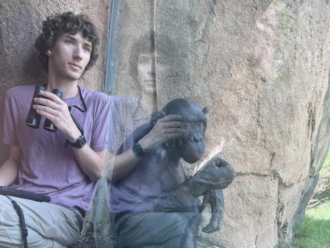
"This photo shows me collecting observational data on the behavior of the bonobos at the Columbus Zoo and Aquarium, where I had the opportunity to watch them nearly every day over the summer of 2025. This involved using my iPad (seen here on my lap) to take careful notes about everything the bonobos did and how they spent their time. One day, I was sitting in the corner using my binoculars to watch a distant group of bonobos (out of frame) when Bila Isia, a 24-year-old male bonobo, walked up to the window and sat down next to me. As our closest living relatives, these apes offer a valuable glimpse into what it means to be human. Just like us, bonobos are highly intelligent and capable of complex emotions, so it’s difficult to watch bonobos without feeling like I’m staring at a reflection of myself and my own humanity."
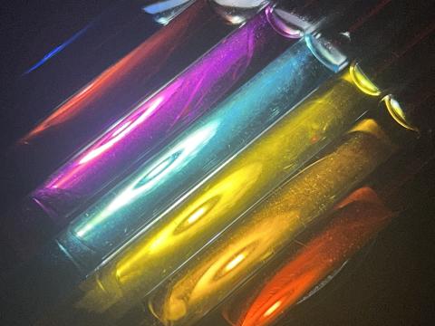
2nd Place: The color of discovery
Image by Natafira Suryanata
"These brilliantly colored NMR tubes hold ruthenium complexes, a family of compounds that has the potential to drastically reduce the necessity of needles in drug delivery. Millions of people worldwide rely on daily injections, but what if a small deposit below the skin could allow patients to self-dose at home simply with a flashlight? This is one of the driving questions being researched in the Rapp Lab. The vibrant colors of these tubes are not only beautiful but informative. Each hue reflects the unique properties of the compound and subtle shifts in their colors inform us of changes happening at a molecular level. This allows us to visually track how these compounds evolve throughout the research process. The Rapp Lab leverages these light sensitive ruthenium compounds to revolutionize bioengineering by targeting, treating, or directing biological events."
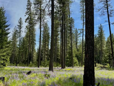
3rd Place: Burning for biodiversity
Image by Flora Booker
"When people ask about the lab I work in, I show them this picture: a ponderosa pine woodland where frequent prescribed burns encourage an abundance of fire-following plants. Thanks to the Vice President for Research and Innovation Fellowship, I am working with the Mount Adams Resource Stewards Prescribed Burn Association, a nonprofit that brings community members together to burn, care for, and learn from the Pine Flats Community Forest. Look closely at the dark burn scars at the base of these trees. Ponderosa pines are fire-adapted, with bark that sheds during burns to help prevent flames from reaching their canopy. On the ground, lupines bloom in bright purples. Their seeds were safely nestled in the soil as fire crept through, then sprouted with the increased nutrients, water, and sunlight left behind. This is my lab, where science meets stewardship!"
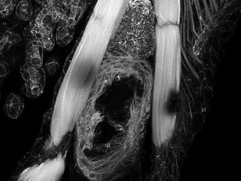
People's Choice: Guard your heart, a lesson from Danio rerio
Image by Sara Swinson
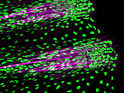
"This cross-sectional view of the heart of a zebrafish focuses on the ventricle and its anterior neighbor: the bulbus arteriosus. The two structures work together to maintain constant flow and blood supply, encouraging our samples to just keep swimming. Made visible through phalloidin, a fluorescent toxin derived from mushrooms, labeled actin filaments illuminate the various protein networks present in the chest cavity. The contrast between the fibrotic cardiac structures and the striated muscles that flank the heart demonstrates the dynamic nature of this protein and the different forms it can take on. Take a minute to appreciate how protected these fish keep their hearts, and try to do the same with yours!"
2025
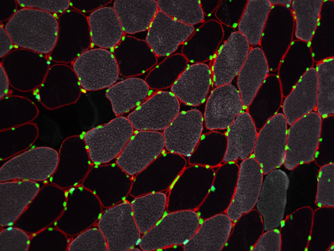
1st Place: A trip down (muscle) memory lane
Image by Kaitlyn Augienello
"A cross-sectional view of human skeletal muscle tissue stained for measurement of various informational parameters. A main goal of our research is to identify trends in muscle across different activity levels over time. This image provides just a momentary snapshot in the storybook of skeletal muscle memory, a phenomena highlighted in our ongoing study. With our analysis, we compile thousands of images and compare the highlighted parameters. This photograph is just the beginning of a Trip Down (Muscle) Memory Lane!"
2nd Place: Red fluorescent protein in C. elegans
Image by Nora Vanasse
"These C. elegans worms have been genetically engineered to express red fluorescent protein, which serves as a vivid biological marker, allowing scientists to track the presence of specific genes in the genome. Fluorescence intensity can qualitatively indicate how much of a particular gene is expressed without any formal genotyping. Fluorescent proteins like RFP are powerful and versitile tools in molecular biology - and they look pretty awesome too!"
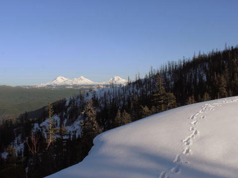
3rd Place: Fire on Carpenter Mountain
Image by Kyla Schmitt
"Last summer, upon receiving the O’Day Fellowship, I got the opportunity to work in the H.J. Andrews Experimental Forest: a long-term ecological research site nestled in the western Cascades, 50 miles from the University of Oregon. The Andrews was swept by wildfire in 2023, simultaneously frustrating ongoing environmental monitoring and creating fascinating natural experiments, facilitating applied research on how post-wildfire conditions and other ecological phenomena interact. Working under my graduate mentor, Ethan Torres, I surveyed vegetation in meter-square quadrats to understand how fungi mediate native plant re-establishment post-burn. After a day of hard work in the field, Ethan treated us to a favorite Andrews view: a lookout from Carpenter Mountain. With a burn-scarred hillside in the foreground and the Three Sisters looming silently in the background, this awe-inspiring landscape instills a strong sense of place, shaping the ecosystems that research teams like ours have the privilege of learning from."
2024
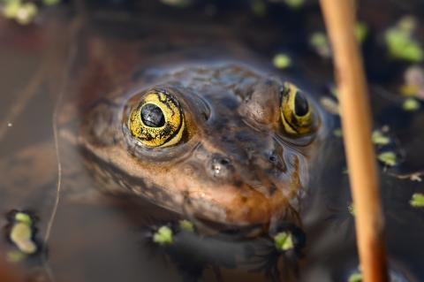
1st Place: Spotting the Oregon Spotted Frog
Photo by Eden McCall
"This photograph I made of the Oregon spotted frog, taken at a small artificial pond in downtown Bend, Oregon, forms a crucial part of my multimedia research project on conservation and science communication. The spotted frog once lived throughout Oregon; however, a now familiar narrative for threatened species, due to habitat loss, invasive species, and climate change, the frog has lost more than 80% of its historic range. Contrary to the notion that it must now live in only remote, pristine areas, the frog is actually found thriving in the heart of a city—between yoga studios and residential complexes—because humans have created a quasi-natural environment that supports it. My work, which spans journalism, spatial data science, and multimedia storytelling, uses immersive photography and other visuals as well as maps and data graphics to show how community-driven conservation efforts support ecosystem health and foster coexistence between humans and nature. This image, capturing the frog in its unexpectedly urban environment and at an intimate range, attempts to connect people with nature and contribute to the narrative about local stewardship and ecological resilience, aiming to inspire awareness and action to protect the natural world."
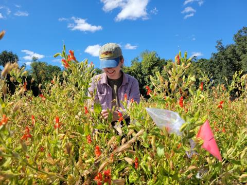
2nd Place: Among the Monkeyflowers
Photo by Olivia Wilborn-Pilotte
"This photo shows me collecting data for my thesis on pollination strategy as an evolutionary response to climate change in a native wildflower species, the scarlet monkeyflower. Here I am using a small glass tube to collect nectar from a flower to measure the volume of nectar and the sugar concentration. The nectar is collected by inserting a small glass tube into the base of the flower, which draws the nectar into the tube via capillary action. The volume is then measured in the tube and the nectar is deposited onto an instrument called a refractometer, which measures the sugar concentration."
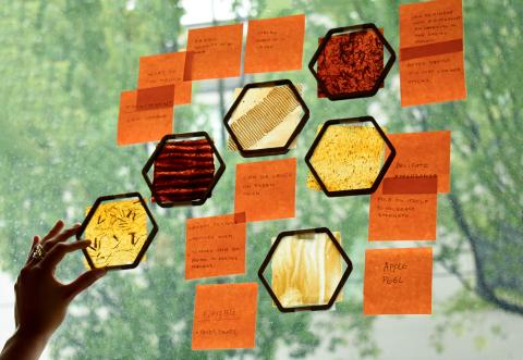
3rd Place: Design for Healing, Restoring Planet Earth
Photo by Tamara Alarcon Basurto
"Documenting the translucent properties of the bio-material composite in natural light, the image showcases the initial stages of my research. Through observation, I recorded my first impressions of the materials I created. Subsequently, I categorized and grouped them based on their similarities in qualities and constraints."